Iwai North America Inc.
541 Taylor Way Suite# 4
San Carlos, CA 94070
Phone : (650) 486-1541
Fax : (650) 394-8638
Open weekdays 9 AM-6 PM (PST)
.........................................................................................................................................................................................................................................................
BioMIMESYS® is an Organ specific Extracellular Matrix (ECM) formed by crosslinking reaction of hydrosoluble modified hyaluronic acid (HA) and other ECM components (collagens, fibronectin, etc.) with ADH (Adipic acid dihydrazide), to create a "Hydroscaffold" 3D cell culture system.
2D cell culture is not a true reflection of the physiological cell environment. In a real body, cells grow in 3D, connecting to other cells and the extracellular matrix (ECM) to build tissues and organs. Therefore, 3D cell culture can better mimic the original tissue’s specific characteristics. Some of the processes studied in 2D culture such as gene expression, apoptosis, and, importantly, drug uptake and toxicity may not be directly transferable to in vivo experiments.
The Importance of ECM
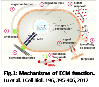
ECM has greatly determines cell activity and viability (1) by:
• Maintaining the structure of tissues and organs
• Defining microenvironment for cells
• Determining cell behavior and function
Changes dynamically through development and function (healthy - diseased). And, notably, ECM composition is organ-specific.
Side Note: Components of ECM
ECM consists of interlocking mesh of fibrous proteins and glycosaminoglycans
Proteins
• Collagen – structural support
• Elastin – provide elasticity
Glycosaminoglycans
• Proteoglycans(GAG) – hydration
• Polysaccharides (hyaluronic acid) - load bearer, hydration
Glycoproteins
• Fibronectin – connect cell with collagen fibers, movement of cells
• Laminin – basal laminae, cell adhesion
Linker proteins such as nidogen and entactin, which connect collagens with other protein components.
The ideal ECM for research is decellularized organs, but the cost, reproducibility, and scalability make it impractical for continuous use. The following characteristics of the ECM that Biomimesys provides makes it the best alternative.
1) BIOMIMESYS is a physiological hydroscaffold to mimic tissue-specific ECM
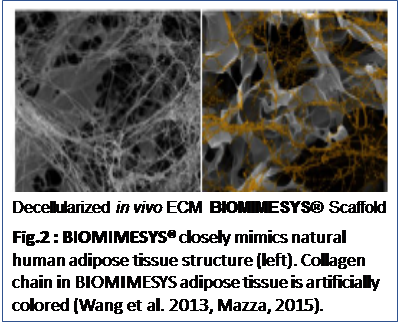
• BIOMIMESYS® scaffold possesses a similar structure and mechanical property in hydrated stage to natural organs (Fig.2)
• Made of high molecular weight hyaluronic acid (1.6 MDa), adipic acid dihydrazide crosslinker, undenatured collagen and other ECM.
• Tissue/organ-specific scaffold available and customizable. Various ECM compositions and physical properties are available and customizable to mimic healthy and diseased tissues.
2) High porosity for greater diffusion
• The porous nature of the scaffold provides greater diffusion of oxygen and nutrients for better viability and greatly decreases the formation of necrotic core, often seen in other spherical 3D models.
• The abundance of metabolic enzyme activities and normal cellular polarity makes it ideal for in vitro models
Side Note:
In addition to oxygen tension, cell behavior in 3D cultures is regulated by a variety of other biophysical and biochemical properties of the microenvironment including CO2 concentration, availability and diffusion properties of nutrients and waste products, matrix rigidity and the presence of matrix-binding factors in the culture medium.
3) Many potential applications and Cells tested
• Microscopy (Immunofluorescence, histology)
• Plate readers (OD, fluorescence & luminescence)
• Protein and RNA extraction, PCR, Western-Blot
References
1.Lu, P., Weaver, V. M. & Werb, Z. The extracellular matrix: A dynamic niche in cancer progression. J Cell Biol 196, 395 (2012).
2.Kola, I. & Landis, J. Can the pharmaceutical industry reduce attrition rates? Nat Rev Drug Discov 3, 711–715 (2004).
3.Wang, L., Johnson, J. A., Zhang, Q. & Beahm, E. K. Combining decellularized human adipose tissue extracellular matrix and adipose-derived stem cells for adipose tissue engineering. Acta Biomater 9, 8921–8931 (2013).Mazza, 2015
4.Mazza, G. et al. Decellularized human liver as a natural 3D-scaffold for liver bioengineering and transplantation. Sci Rep 5, 13079 (2015).
5.Lodish, H. et al. Integrating Cells Into Tissues. in Molecular Cell Biology (Freeman, W. H. & Company, 2003).
6.Peach, R. J., Hollenbaugh, D., Stamenkovic, I. & Aruffo, A. Identification of hyaluronic acid binding sites in the extracellular domain of CD44. J Cell Biol 122, 257–264 (1993).
7. Colom, A. et al. Oxygen diffusion and consumption in extracellular matrix gels: implications for designing three-dimensional cultures. J Biomed Mater Res A 102, 2776–2784 (2014).
8.McMurtrey, R. J. Analytic Models of Oxygen and Nutrient Diffusion, Metabolism Dynamics, and Architecture Optimization in Three-Dimensional Tissue Constructs with Applications and Insights in Cerebral Organoids. Tissue engineering. Part C, Methods 22, 221–49 (2016).
9. Al-Ani, A. et al. Oxygenation in cell culture: Critical parameters for reproducibility are routinely not reported. PLoS One 13, e0204269 (2018)
Why 3D Cell Culture?
Characteristics of hyaluronic acid
• Main component of the interstitial gel.
• Found on the inner surface of the cell membrane and translocate out of the cell during biosynthesis.
• Known to be less toxic and cause less allergic reaction. Can be used for in vivo experiment models.
• Absorb significant amount of water to allowing tissues to swell thus enabling it to resist compression.
• Acts as an environmental cue that regulates cell behavior during embryonic development, healing processes, inflammation, and tumor development.
• Interacts with a specific transmembrane receptor, CD44 (Ref: Lodish et al, Peach 1993).
Ideal 3D Models - Why BIOMIMESYS
Fig.1: Mechanisms of ECM function.
Lu et al. J Cell Biol. 196, 395-206, 2012
Ready to Use
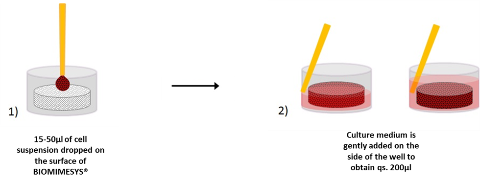
BIOMIMESYS® is EASY & READY TO USE. Upon receiving the vacuum sealed 96-well plate, open it (under a hood) and add the cells directly on top of the matrix. Changing the culture medium is easy as well. To remove medium, simply draw the medium with a pipette between the matrix and the edge of the well. To refresh the medium, place fresh medium onto the surface of the matrix.
Request a Free Sample
Please contact us for more information about BIOMIMESYS samples. A representative from our sales team will be in contact with you as soon as possible.
Cells Tested
Click Here for a list of cells tested. Please note that new cells are always being tested. Please email us at orders@iwai-chem.com with the name of the cell, if you do not see it in the list.

